68Ga-DOTA-Siglec-9 PET Imaging Detects Biomaterial Infection Caused by S. Epidermidis
Authors: Aro HT, Ahtinen H, Kulkova J, Moritz N, Lindholm L, Eerola E, Hakanen A, Söderström M, Saanijoki T, Roivainen A. University of Turku, Turku, Finland
Title: 68Ga-DOTA-Siglec-9 PET Imaging Detects Biomaterial Infection Caused by S. Epidermidis
Background: AAOS gave only a weak recommendation for nuclear imaging in the diagnosis of periprosthetic infections. Reflecting limited inflammatory reaction, subacuteperi-implant S. epidermidis infections were characterized by low 18F-FDG uptake in a recent experimental study of 18F-FDG-PET/CT. Vascular adhesion protein-1 (VAP-1) is an inflammation inducible endothelial protein. Siglec-9 is a leukocyte ligand of VAP-1. 68Ga-labeled Siglec-9 is a potential novel tracer for PET imaging of low-grade S. epidermidis infections.
Hypothesis/Purpose: We asked whether the 68Ga-DOTA-Siglec-9 PET/CT imaging is able to (1) detect aseptic inflammation in a bone and (2) to differentiate peri-implant infections caused by S. epidermidis or S. aureus in comparison with aseptic inflammation.
Methods: Thirty adult Sprague-Dawley rats were randomized into three groups (n=10/group). A polymer catheter was placed into the medullary cavity of the left tibia followed by injections of S. epidermidis (T-54580, 3 × 10⁸ CFU/mL) or S. aureus (52/52A/80, 3 × 10⁵ CFU/mL) with an adjunct sodium morrhuate. In the negative control group, equal amount of sterile saline was injected via the catheter. The catheter, cut at the level of tibial tuberosity, was left in situ to serve as the implant. At two weeks, PET imaging with 68Ga-DOTA-Siglec-9 was performed with quantitative analysis of the standardized uptake value (SUV). The presence of infections and the absence of bacterial contamination in the control group were verified by microbiological analyses. Histologic inflammatory reaction was graded using a scoring system.
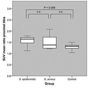
Results: There was a significant difference (29.5%, p<0.001) in the uptake (SUV ratio) between the operated and contralateral tibia in the control group. The corresponding SUV ratio values were 58.1% in the S. epidermidis group and 41.7% in the S. aureus group. The uptake was significantly (p=0.009) higher in the S. epidermidis group than in the control group (Figure). The difference between the S. epidermidis and S. aureus groups was not significant.
Discussion: The animal model was reproducible in creation of culture-positive implant-related infections. As a limitation, S. epidermidis infections were moderate/severe and not low-grade subacute infections.
Conclusion: 68Ga-labeled Siglec-9 PET/CT imaging demonstrated aseptic inflammation in bone in response to catheter implantation. The method also detected peri-implant infections caused by S. epidermidis